Our Research

Exploring the frontiers of cellular signaling and molecular dynamics

Ongoing Projects

Regulation of Raf activity by KRas dimerization dynamics
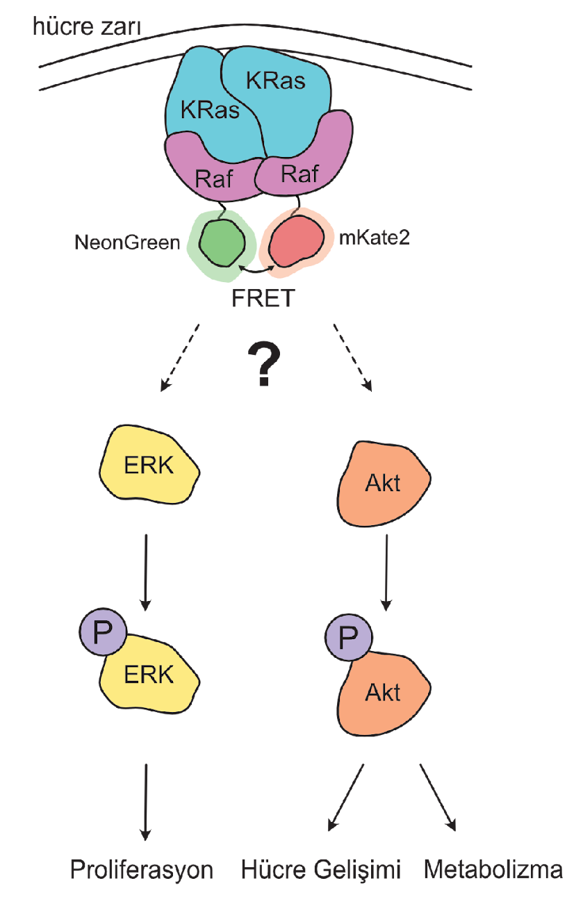
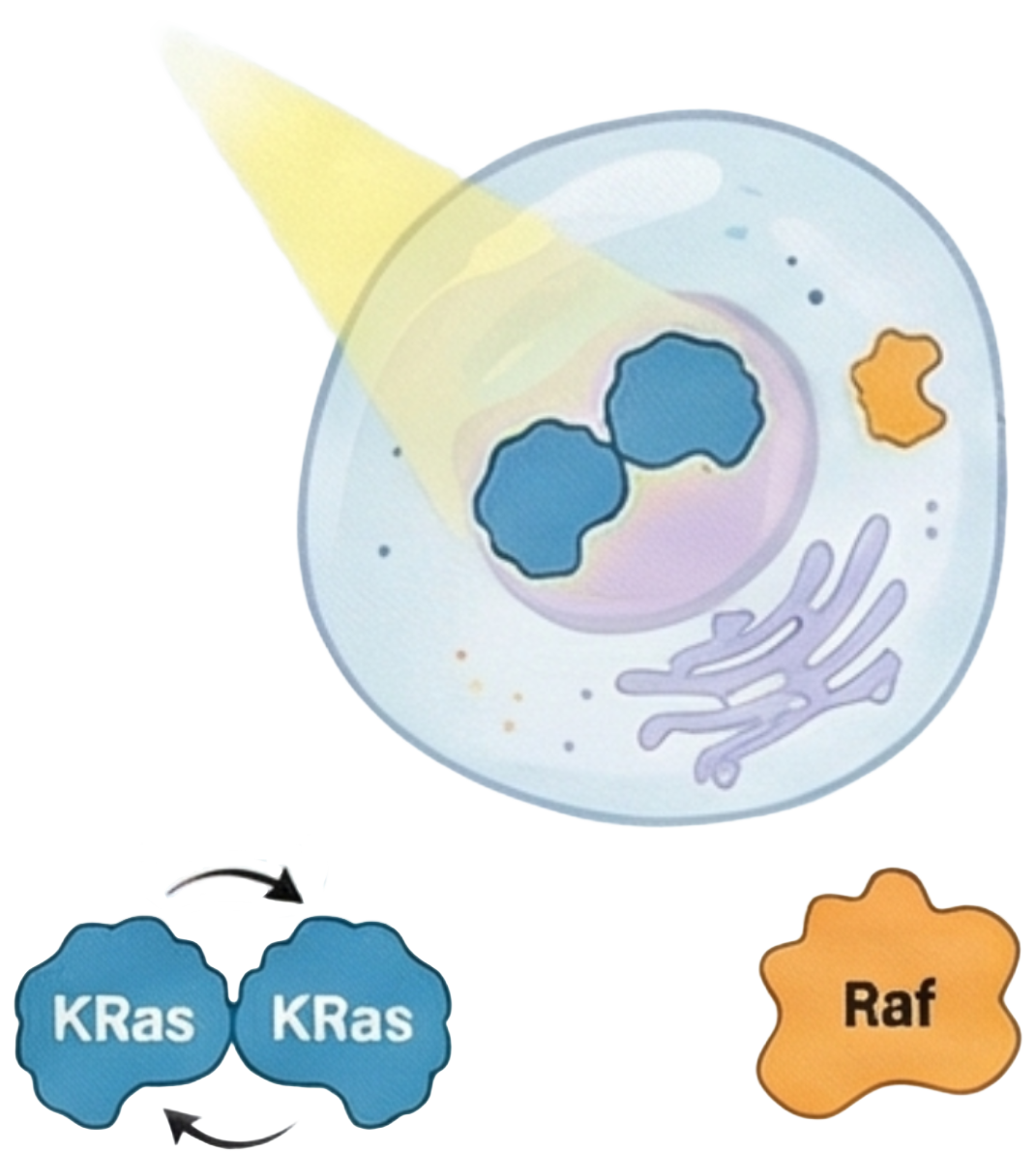
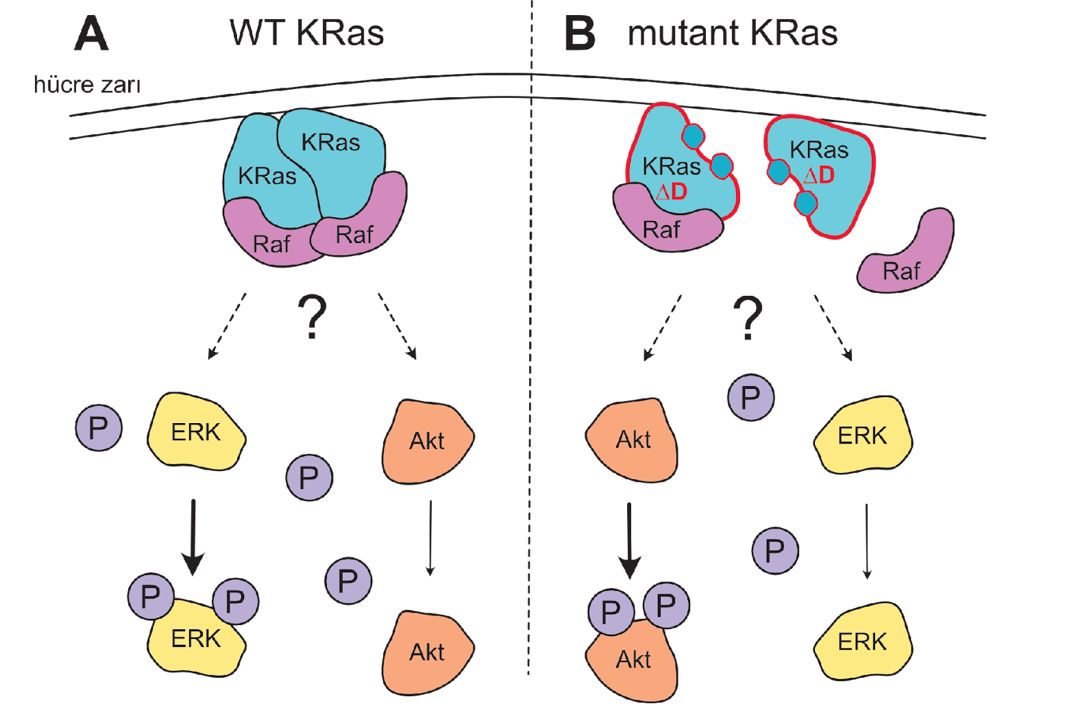
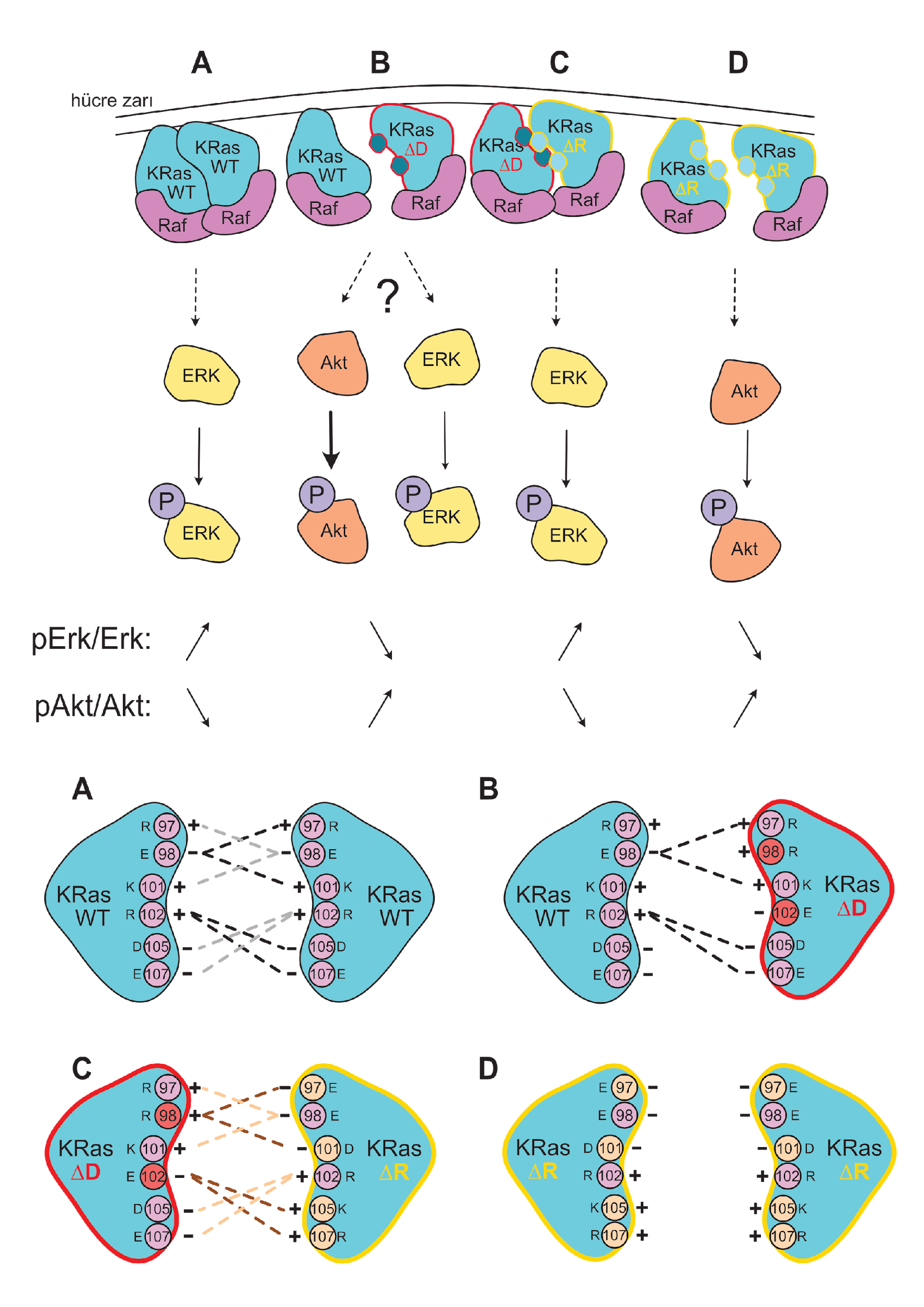
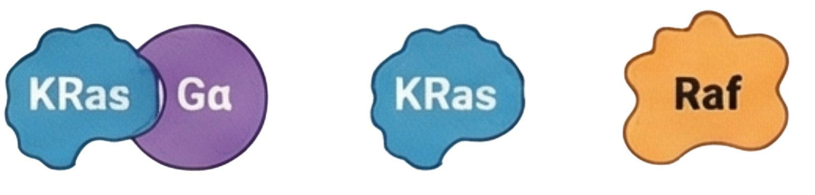
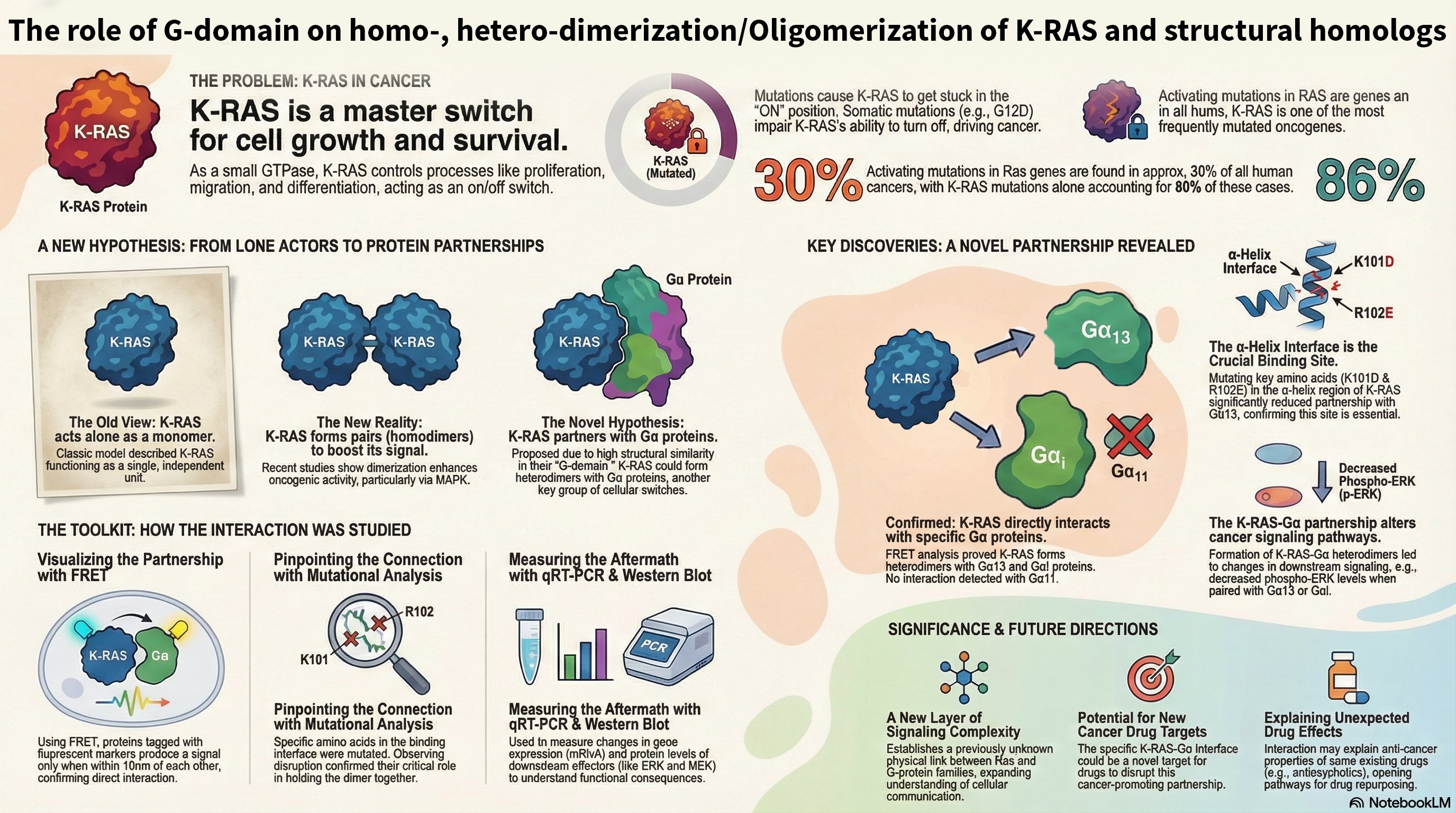
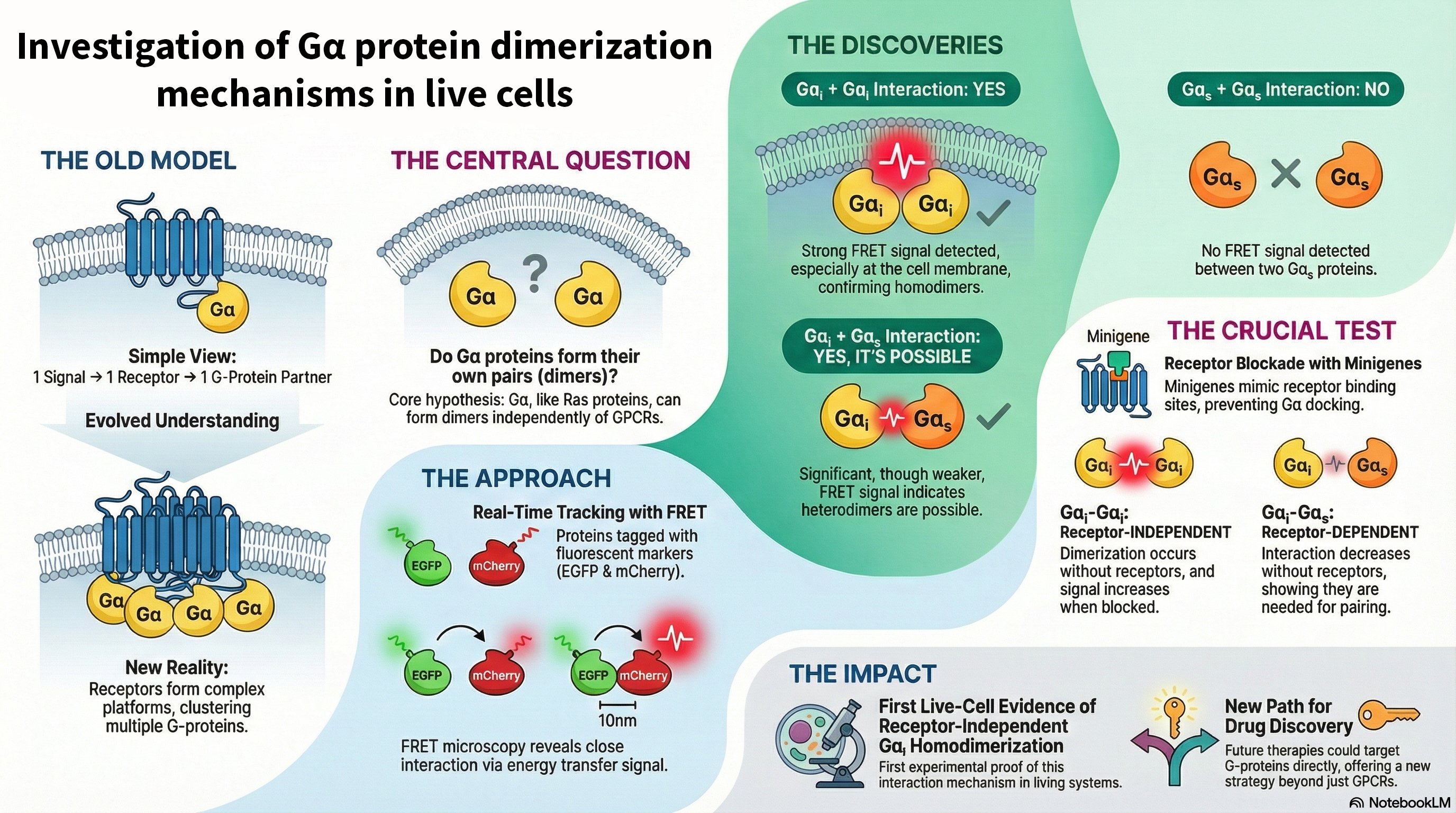
Overview: KRas is a critical GTPase in cellular signal transduction, regulating key pathways like MAPK/ERK and PI3K/AKT through its interaction with effectors such as Raf kinase. This project investigates the fundamental mechanism of Raf activation, specifically questioning whether KRas homodimerization is essential or if monomeric KRas is sufficient to drive downstream signaling. We explore the dynamics of KRas dimerization, potential compensatory mechanisms involving KRas-Gα heterodimerization, and how various KRas mutations influence signaling outcomes in both cancer and neuronal contexts.


Key Research Areas:

  • Dimerization vs. Monomeric Activation: Using live-cell imaging and Förster resonance energy transfer (FRET), we are dissecting whether KRas monomers can activate Raf independently or if dimerization is required for a stable, prolonged signaling response.
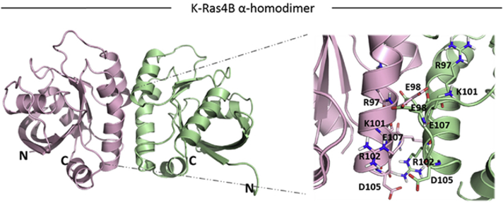
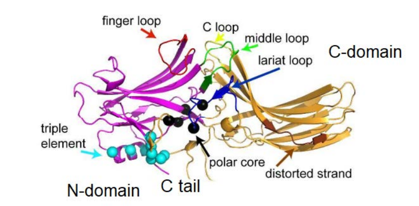
  • Mutational Analysis of the Dimerization Interface: We employ precisely designed KRas mutations at key residues (e.g., Glu98, Arg97, Arg102) to selectively disrupt or rescue homodimerization. This genetic approach allows us to isolate the specific role of dimerization in signaling, a significant advantage over traditional chemical inhibitor methods.
  • Compensatory Mechanisms: We are investigating whether KRas-Gαi/Gα13 heterodimerization can serve as an alternative pathway to sustain Raf activation when homodimerization is compromised, potentially revealing novel cross-talk between GPCR and Ras signaling pathways.

Significance and Implications: By understanding the molecular nuances of KRas-driven signaling, this research aims to identify new regulatory nodes for therapeutic intervention in cancer, where KRas mutations are prevalent. Furthermore, given KRas's role in neuronal differentiation and synaptic plasticity, our findings could offer new insights into neurodevelopmental disorders and neurodegenerative diseases.

Figure 1: KRas Dimerization Model
Fig 1: Model of KRas dimerization at the plasma membrane and its role in Raf activation.
Figure 2: FRET Imaging Data
Fig 2: KRas interaction dynamics in live cells.
Figure 3: Signaling Pathway Diagram
Fig 3: Diagram illustrating the downstream effects of KRas signaling on the MAPK/ERK and PI3K/AKT pathways.

The Effects of K-Ras/G-alpha Heterodimerization on Raf Activation

Overview: Raf proteins are central to the MAPK signaling pathway, but the precise mechanism of their activation by K-Ras remains debated. While Ras dimerization is known to be a key step, conflicting findings exist regarding the necessity of K-Ras homodimerization for Raf activation. This project hypothesizes that these discrepancies arise from overlooking the potential role of K-Ras forming heterodimers with structurally similar Gα proteins.


Key Objectives:

  • Real-Time Observation: To directly observe the interaction between K-Ras and Raf dimers in real-time within live cells using FRET, providing clear evidence of the molecular dynamics at play.
  • Isolating Heterodimer Effects: To engineer a novel model system using mutant K-Ras and Gα proteins. These mutants are designed to inhibit homodimerization while promoting heterodimerization, allowing us to isolate and study the specific effects of K-Ras/Gα pairs on downstream signaling.
  • Investigating Raf Activation: To use our model system to determine whether K-Ras/Gα heterodimers have a positive or negative regulatory effect on Raf dimerization and activation, clarifying their role in the signaling cascade.

Significance and Implications: This research aims to resolve a key controversy in cell signaling and uncover a novel regulatory mechanism involving Gα proteins. Understanding this K-Ras/Gα cross-talk could have significant implications for both cancer biology and neurology, potentially revealing why certain drugs for neurological disorders might have unexpected effects in K-Ras-driven cancers and paving the way for new therapeutic strategies.

Figure 1: K-Ras and G-alpha Heterodimerization Model
Fig 1: Proposed model of K-Ras/Gα heterodimerization influencing Raf activation.

Automation of Confocal Microscope Imaging and Data Processing

Overview: Manual confocal imaging is time-consuming and prone to researcher-to-researcher variability. This project aims to integrate a standardized, autonomous control system into our existing confocal microscope and to develop in-house Python software for fully automated image acquisition and processing.


Key Research Questions

  • Can confocal microscope imaging be automated and standardized to eliminate human error?
  • Will automated image processing deliver higher efficiency and reproducibility than current manual methods?

Methodology

  • Hardware Automation: Design and fabricate an x-y-z motorized stage and integrate control electronics to retrofit the current microscope.
  • Control Software: Develop algorithms for autonomous stage movement, autofocus and image stitching to create seamless large-area views.
  • Image Processing Pipeline: Implement Python scripts for ROI detection, denoising and FRET-ready image generation; compile resulting data sets for downstream analysis.
  • Machine Learning: Feed processed images into neural-network classifiers to discover interaction-specific patterns.

Expected Outcomes

  • Fully automated x-y-z stage control enabling high-throughput imaging.
  • Standardized processing pipeline that converts raw images into analysis-ready data sets.
  • Publicly available interaction-specific image databases for the research community.
  • Proof-of-concept ML models that classify protein-interaction images with high accuracy.

Investigation of the Effect of SPOCK1 Protein on Apoptosis in Nerve Cells

Collaborative Project

Overview: With the development of health services, the elderly population is increasing around the world, leading to a rise in age-related diseases. Late-onset Alzheimer's disease (LOAD) occurs in eight out of 100 people over the age of 65. The accumulation of amyloid plaques in the brain and neurofibrillary tangles (NFT) formed by tau proteins are important molecular markers of Alzheimer's disease (AD). These molecules contribute to the formation of reactive oxygen species (ROS) by causing inflammation.


Key Research Areas:

  • SPOCK1 in Mitochondria: Investigating the presence and role of SPOCK1 in mitochondria of astroglia and neuroblastoma cells, despite being known as a secreted protein.
  • Apoptosis Mechanisms: Examining the relationship between SPOCK1 and apoptosis in nerve cells, particularly in the context of AD-related cell death pathways.
  • ERBB4 Pathway: Exploring the potential regulation of SPOCK1 via the ERBB4 pathway in different types of nerve cells.
  • Cell Type Specificity: Comparing the effects of SPOCK1 across astroglia, microglia, and neuroblastoma cells to understand cell-type specific responses.

Significance and Implications: This research aims to uncover the role of SPOCK1, a member of the SPARC family, in Alzheimer's disease progression. By understanding how SPOCK1 influences apoptosis in nerve cells and its potential regulation through the ERBB4 pathway, we may identify novel therapeutic targets for AD treatment. The project could provide crucial insights into the molecular mechanisms underlying neurodegeneration and open new avenues for intervention strategies.

Figure 1: SPOCK1 in Alzheimer's Disease
Fig 1: Basic flow of the development of Alzheimer's disease.
Figure 2: SPOCK1 and Beta-Amyloid Interaction
Fig 2: Downstream targets of ERBB4.
Figure 3: SPOCK1 Expression in AD Brain
Fig 3: Proposed flow of the project.

Activation of Arrestin-mediated signaling pathways by small molecules

Collaborative Project
Primary Investigator: Özge Şensoy, Assoc. Prof. Dr.

Overview: The discovery of initiation of G protein-independent signaling pathways by Arrestin, which is responsible for termination of GPCR-signaling, has been the breakthrough in the field of GPCR. This has led to development of novel class of ligands, namely biased ligands, which can provide specific coupling of the receptor either to G protein or Arrestin. Sometimes, Arrestin signaling pathways provide advantages over G protein-mediated signaling pathways.


Key Research Areas:

  • Structural Modeling: Building 3D models of phosphorylated GPCR/ligand/Arrestin complexes to understand the molecular interactions at atomic resolution.
  • Molecular Dynamics Simulations: Performing simulations in physiologically relevant membrane environments to determine Arrestin conformations and dynamics.
  • Small Molecule Discovery: Identifying and characterizing small molecules that can stabilize specific Arrestin conformations for therapeutic applications.
  • Machine Learning Approach: Developing computational methods to predict and design ligands that directly bind to and modulate Arrestin activity.
  • Experimental Validation: Testing candidate molecules using BioID method to identify Arrestin-interacting proteins and validate their functional effects.

Significance and Implications: This research aims to test the hypothesis that signaling proteins determined by GPCR-bound ligands can be targeted by small molecules that directly bind to Arrestin. Using β2-adrenergic receptor (β2AR), Arrestin 3, and both biased (carvedilol) and classical (isoproterenol) ligands as a model system, this project will provide crucial insights into the molecular mechanisms of biased signaling. The findings could lead to novel therapeutic strategies for conditions where GPCR signaling is dysregulated, such as cardiovascular diseases and potentially COVID-19 related respiratory complications.

Collaboration: Computational studies led by Assoc. Prof. Dr. Ozge Sensoy (Istanbul Medipol University), method development by Dr. Giulia Morra (Italian National Research Council), and experimental studies supervised by Assoc. Prof. Dr. Cagdas Devrim Son (METU).

Figure 1: Arrestin-mediated signaling
Fig 1: Arrestin-mediated signaling pathways and their therapeutic potential (Mohad et al., 2012).
Figure 2: 3D Structure of Arrestin
Fig 2: Three-dimensional structure of Arrestin showing potential binding sites.
Figure 3: Arrestin Conformational Changes
Fig 3: Conformational changes in Arrestin upon receptor binding (Ostermaier et al., 2014).
Figure 4: Biased Ligand Effects
Fig 4: Differential effects of biased ligands on Arrestin-mediated signaling (Rajagopal et al., 2011).

Completed Projects

Information on completed projects will be available soon.